Preanalytical factors have a negative impact on sample quality
If you’re working with blood sample collection, you might have heard the term ‘preanalytical factors’ at some point. Then again, you might not. In many studies, preanalytical factors are almost completely ignored. So what are they? Preclinical factors are all aspects of a sample preparation protocol that affect the outcome of sample analysis. For example, if you’re collecting and preparing a blood sample for flow cytometry analysis, preclinical factors would include things like the anticoagulant used, the storage time and temperature of the blood sample before processing, the processing protocol used, and the mode of storage of the purified cells.
At this point, there is a large body of evidence demonstrating the negative impact of preanalytical factors on clinical studies, but how do they arise? Immediately after a blood sample is collected, a deterioration process sets in. The blood cells start consuming plasma metabolites and nutrients, change their gene transcription programs to accommodate their new milieu, and eventually, they will start dying. If a blood sample is not processed immediately, it will lead to changes in antigen expression (1), gene expression (2), protein secretion (3), cell viability (4), and cell counts (5).
The trouble doesn’t end there. The protocols for the isolation of white blood cells include exposing the cells to harsh chemicals that induce various stress responses in the cells and make it harder to preserve the original phenotype they exhibited in vivo. Red blood cell lysis protocols affect the cell count (6), cell type frequencies (7–9), and surface antigen expression (9) of the sample. The popular cell preparation method, density gradient centrifugation, or ‘Ficoll-prep’, is also detrimental to the sample and induces changes in cell type frequencies (10), surface antigen expression (11), gene expression (12), and protein production (13). Finally, cryogenic preservation of the isolated cells also changes the cell counts, viability, frequency, antigen expression, gene expression, and protein secretion of the preserved sample (14, 15).
Stressful clinical environments make sample preparation difficult
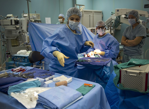
It is clear that preclinical factors influence sample quality and that these changes affect the study outcome (16). So why do these changes occur? The current gold standard blood processing methods are complicated, so delays due to practical considerations at the sampling site are common, especially in stressful and low-resource clinical environments. The protocol details often differ between studies, and the many steps increase the risk of mistakes and operator bias. Most importantly, manipulation-induced cellular changes are simply inherent in the methods and won’t go away even if the protocols are followed stringently.
We have made it our mission to bring clinical sampling technology into the 21st century. Our improved solutions for blood sample preparation make blood sampling far easier and cheaper than older methods and increase data quality. Our Whole Blood Stabilizer Buffer enables on-site blood preservation with only a single mixing step. The white blood cells are stabilized immediately after sampling, minimizing the effect of preanalytic factors. Our WB-STIM blood stimulation system allows for functional immune testing, which has been proven to be more physiologically relevant and reproducible than the stimulation of purified cells (17, 18).